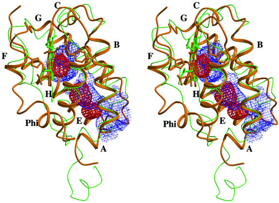
Figure 2.
Stereo view of the trHbO subunit tertiary structure (orange ribbon) overlaid onto trHbN (green trace). Individual α-helices are labeled for trHbO. The figure includes mesh surfaces defining the trHbO protein matrix cavities (red color), and the matrix tunnel identified in trHbN (blue color).

