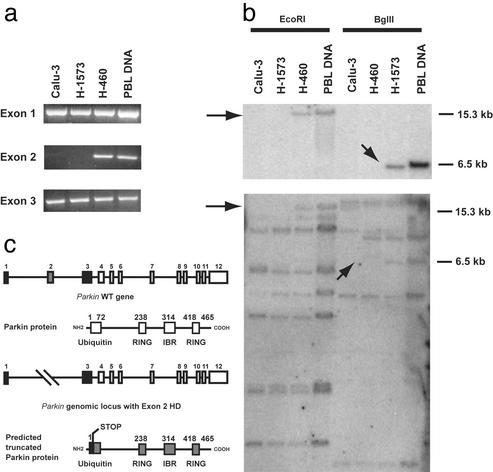
Figure 4.
Analysis and mapping of HDs in the Parkin locus. (a) PCR analysis of exons 1–3 in Calu-3, H-1573, and H-460 cell lines and in normal human genomic DNA. (b) Southern analysis of Parkin gene in DNA digested with EcoRI or BglII. Blots were hybridized with either a Parkin exon 2-specific probe (Upper) or the full-length Parkin cDNA (Lower). Arrows indicate the absence of the expected 15.3- and 6.5-kb EcoRI and BglII fragments, respectively. PBL, human peripheral blood leukocyte. (c) A schematic representation of exon 2 deletions observed in the lung adenocarcinoma cell lines, Calu-3 and H-1573.

