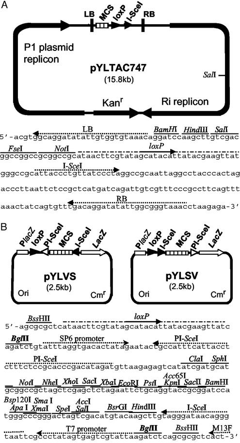
Figure 1.
Physical maps of the acceptor vector pYLTAC747 and donor vectors pYLVS and pYLSV. (A) Structural features of the pYLTAC747 and sequence between the T-DNA LB and RB. Four unique restriction sites, a loxP site, and a recognition site for I-SceI were placed between LB and RB. The plasmid backbone contains a kanamycin-resistance gene (Kanr, modified NPTI), a P1 plasmid replicon for replication in E. coli, and an Ri replicon for replication in Agrobacterium. (B) Structural features of the donor vectors and a part of sequence of pYLVS showing the key sites. The orientation of the segment between the two BglII sites is reversed in the two constructs. Cmr denotes the chloramphenicol-resistance gene and Ori denotes the ColE1 replicon.

