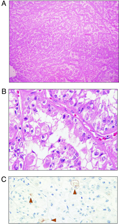
Figure 1.
Light microscopic features of t(6;11) carcinoma. (A) At low magnification, the tumor consisted of epithelioid cells arranged in a nested alveolar or acinar pattern (H&E, original magnification, ×5). (B) Higher magnification showed cells with voluminous clear and granular eosinophilic cytoplasm. Acinar lumens often contained clusters of degenerating cells with pyknotic nuclei. Individual cells were round to polygonal with abundant cytoplasm. Acinar structures were separated by thin vascular channels (H&E, original magnification, ×60). (C) Immunostaining showed rare focal positivity for HMB-45 (H&E, original magnification, ×40).

