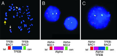
Figure 3.
(A) 6p21.1 translocation breakpoint evaluated in t(6;11)(p21.1;q13) metaphase cell by dual-color FISH. BACs telomeric (red) and centromeric (green) to TFEB are separated by the translocation. Yellow arrow indicates overlapping signal from nontranslocated chromosome. (B) 11q13 translocation breakpoint evaluation by using BACs telomeric (red) and centromeric (green) to the Alpha locus. Rearrangement of this locus is shown in each of four interphase tumor cells. (C) Colocalization of TFEB centromeric BAC (green) and Alpha telomeric BAC (red) in tumor interphase cell. Overlapping green and red FISH signals appear yellow when merged.

