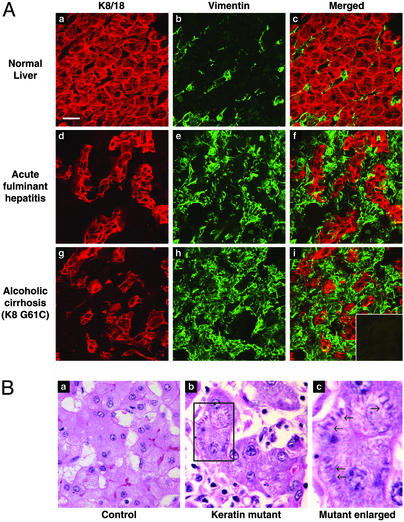
Figure 2.
Keratin filament organization in human liver explants, and histologic findings of livers harboring the keratin mutations. (A) Human livers were sectioned, fixed in acetone, and double-stained with rabbit anti-K8/18 (red) or mouse anti-vimentin (green) antibodies. (i Inset) Control double staining using red and green fluorochrome-conjugated goat anti-rabbit and goat anti-mouse antibodies without adding any primary antibodies. For each tissue, the “Merged” image corresponds to K8/K18 plus vimentin staining. All images were obtained by using the same magnification. (Bar in a = 20 μm.) (B) Hematoxylin and eosin staining of explanted liver from two patients with acute fulminant hepatitis. a is from a patient without a keratin mutation, whereas b is from a patient with the K18 T102A mutation. The region outlined by a box in b is magnified in c to illustrate the cytoplasmic filamentous deposits noted primarily in livers of patients with keratin mutations.

