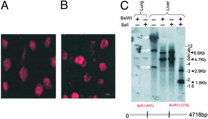
Figure 4.
Molecular characterization and cellular distribution of AAV sequences in tissues. (A and B) In situ hybridization was performed on liver sections from rhesus macaques by using a digoxigenin-labeled AAV8 probe, and optical sections were collected at 0.5-μm intervals. A digital merge of individual green (digoxigenin) and red (propidium iodide) channel images from midplane sections of animals with >20 (RQ4407, A) and <0.1 (V383, B) proviral copies per diploid genome is shown. (C) Molecular state of AAV sequences in the cellular DNA from liver and lung tissues of rhesus macaque RQ4407 was analyzed by DNA hybridization. Total cellular DNA was analyzed by DNA hybridization as described in Materials and Methods. The enzymes used are indicated, and the probe was a mixture of Rep and Cap sequences.

