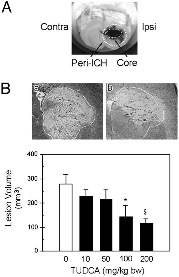
Figure 1.
Collagenase induces intrastriatal hemorrhage in rats. (A) Coronal section of the brain 2 days after ICH. The ipsilateral (Ipsi) ICH core and its periphery (Peri-ICH) are outlined. The contralateral (Contra) hemisphere was analyzed as control. (B) The total lesion volume was markedly reduced in animals receiving TUDCA (10–200 mg/kg of bw) given 1 h before collagenase. Representative Nissl-stained striatal sections are shown for each treatment group. The vehicle lesion (a) is outlined for reference and superimposed on the striatum of TUDCA animals (b). Quantitation of lesion volumes is the mean ± SEM (n = 5 animals per group). *, P < 0.05; §, P < 0.01 from vehicle.

