Figure 2.
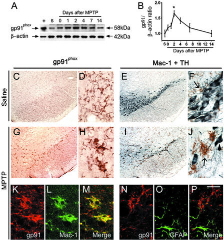
Western blot shows the time-dependent induction of gp91phox in mouse ventral midbrain after MPTP injections. +, Mouse macrophage lysate; s, saline. In saline-injected mice, gp91phox immunoreactivity (C and D, brown) is mild and localized in resting microglia, which are not abundant in the SNpc, as shown (E and F) by Mac-1 labeling (brown, arrow) and are intermingled with TH-positive neurons (gray-blue). Two days after MPTP injections, numerous gp91phox-positive cells are seen in the SNpc (G and H). These cells resemble activated microglial cells (H vs. J, arrow). At this point there are many fewer TH-positive neurons (I and J, arrowhead). Confocal microscopy shows that all gp91phox-positive cells are Mac-1-positive, thus confirming their microglial origin (K and L). Conversely, no gp91phox-positive cells are glial fibrillary acidic protein-positive cells, thus excluding their astrocyctic origin (N–P). *, P < 0.05, more than saline-treated mice (n = 6 per time point). [Scale bar, 2.5 mm (C, E, G, and I); 0.25 mm (D, F, H, and J); and 0.2 mm (K–P).]

