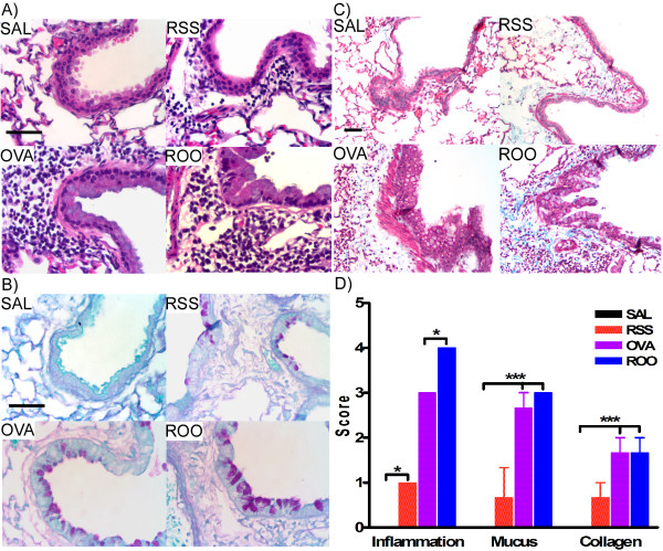
Figure 5.
Lung histopathology of mice exposed to RSV and/or Ova. Lung tissue sections were obtained from mice on protocol day 69. Tissue sections were stained with H&E (A), PAS (B), and MT (C). A. Lung inflammation, B. mucus (purple) hyperproduction, and C. collagen (blue) deposition were observed in mice exposed to RSV and/or Ova. D. Scores were assigned to these histological endpoints by two independent observers and were recorded on a scale of 0–4. Increased deposition of peribronchial and perivascular collagen was observed in the subepithelial reticular layer of the airways in RSS, OVA and ROO mice. In all cases, neonatal RSV infection (RSS) induced persistent lung pathologies including increased peribronchial inflammation, mucus production, and subepithelial fibrosis were exacerbated by subsequent exposure to allergen (ROO). n = 3/group. ***p < 0.001, *p < 0.05.

