Figure 3.
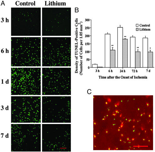
Lithium suppresses MCAO/reperfusion-induced DNA damage in rat brain. Rats were subjected to MCAO for 1 h followed by reperfusion for the indicated times. LiCl (1 mEq/kg) or normal saline was injected immediately after MCAO and daily thereafter as indicated. Brains were sliced with a cryostat into sections of 10-μm thickness and evaluated by TUNEL assay. The sections were also double-stained with NeuN. (A) Representative time-dependent changes in TUNEL staining in a defined locus within the cortical ischemic penumbra area of saline- and lithium-treated rats. (B) Quantification of the density of TUNEL-positive cells shown in A. (C) At 72 h after the ischemic insult, a great majority of TUNEL-positive cells (green) were colabeled with NeuN (red) in the ischemic penumbra area. Bar graphs shown are means ± SEM of TUNEL-positive cells from four rats in each group. *, P < 0.05; **, P < 0.01, compared with the corresponding saline-control group. (Bar: 100 μm.)

