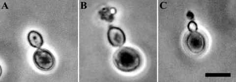
FIG. 3.
Pore formation in yeast cells by scFv antibodies shown by phase-contrast microscopy of S. bayanus AKU 4103 cells treated with scFv-A2 and HM-1. The sample preparation is described in Materials and Methods. (A) Control yeast cells; (B) HM-1-treated cells; (C) scFv-A2-treated cells. Bar, 5 μm.

