FIG. 3.
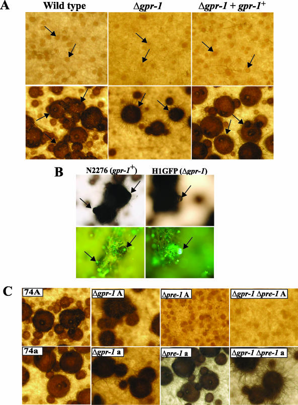
Phenotypic characterization during the sexual cycle. (A) Unfertilized (protoperithecia) and fertilized (perithecia) structures. Strains were cultured on solid SCM at 25°C for 6 days in light (top panels; protoperithecia). At this time, half of the plate was fertilized with wild-type conidia of opposite mating type (74a or 74A) and photographed 3 days after fertilization (bottom panels; perithecia). For the analysis, wild-type (74A), Δgpr-1 (28-6 and 20-1), and Δgpr-1 + gpr-1+ (R28-6) strains were used. Arrows indicate protoperithecia (top panels) or perithecia (bottom panels; enlarged dark bodies). Photographs were taken at ×25 magnification. (B) Microscopic images of perithecial beaks. Strains expressing a histone H1-GFP fusion protein, N2276 (gpr-1+ strain) and H1GFP (Δgpr-1 strain), were cultured on SCM plates for 6 days and then fertilized with opposite mating type conidia. Seven days later, perithecia were subjected to phase-contrast microscopy (upper panels) and detection of GFP fluorescence (bottom panels) (see Materials and Methods). Images are shown at ×200 magnification. The arrows indicate perithecial beaks (left panels) or ruptured perithecia (right panels). (C) Epistasis analysis between gpr-1 and pre-1. Strains were cultured to produce perithecia as indicated in panel A. Wild-type mat A (74A) and mat a (74a), Δgpr-1 mat A (28-6; Δgpr-1A), Δgpr-1 mat a (20-1; Δgpr-1 a), Δpre-1 mat A [(16)A; Δpre-1 A], Δpre-1 mat a [(16)a; Δpre-1 a], Δgpr-1 Δpre-1 mat A (GR1PRE1A; Δgpr-1 Δpre-1 A), and Δgpr-1 Δpre-1 mat a (GR1PRE1a; Δgpr-1 Δpre-1 a) strains were used for analysis. Photographs were taken at ×25 magnification.

