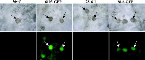
FIG. 5.
Localization of a GPR-1-GFP fusion protein. Phase-contrast (upper panels) and GPR-1-GFP localization (bottom panels) images from 6-day-old unfertilized SCM tissues were obtained as described in the legend of Fig. 3B. Strains carrying gpr-1+ (his-3) and Δgpr-1 (28-6-1) untransformed controls or those expressing a GPR-1-GFP fusion protein in the gpr-1+ (6103-GFP) or Δgpr-1 (28-6-GFP) strain background were used for analysis. Images are shown at ×200 magnification. Arrows indicate protoperithecia.

