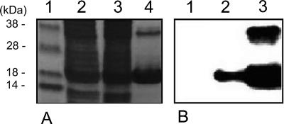
FIG. 8.
In vitro secretion analysis of X. oryzae pv. oryzicola expressing Hpa1. Purified proteins from disrupted cells and the culture supernatant were analyzed by 12.5% SDS-PAGE (A) and immunoblotted with the monoclonal antihexahistidine antibody (B). The plasmid pUhpa1 was used to express the His-tagged Hpa1 in bacteria that were cultured in XOM3 for 16 h. Lanes 1, disrupted cells of the hrpX mutant RCX harboring pUhpa1; lanes 2, disrupted cells of the wild-type RS105 harboring pUhpa1; lanes 3, supernatant of the wild-type RS105 with plasmid pUhpa1. The Hpa1 monomer and dimer are shown as the lower and upper bands in lanes 3. The first lane in panel A is the protein marker.

