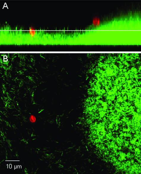
FIG. 1.
Micrograph of C. parvum oocysts (red) associated with a P. aeruginosa (PDO300) biofilm (green). C. parvum oocysts are labeled with a Cy3-conjugated monoclonal antibody solution specific for Cryptosporidium, and P. aeruginosa cells are chromosomally tagged with a green fluorescent protein. (A) Sagittal view of the biofilm. (B) Planar view of the biofilm at the depth indicated by the white line in the sagittal view.

