
FIG. 6.
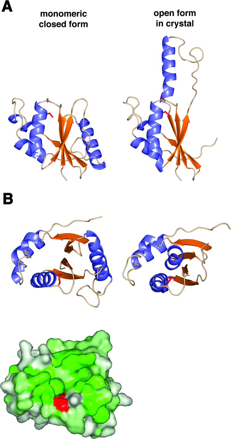
Model of monomeric G4. (A) A monomeric, closed model of G4 (left) was built based on the location of β2b and α2 in the dimer. For reference, the open form of the dimer in the crystal is shown on the right. (B) A view oriented from the top as in Fig. 5B shows the closed model (left) and open model (right) of G4 as a ribbon diagram (above). The same view of the closed model as a surface representation colored by hydrophobicity shows a similar hydrophobic cleft above the active-site cysteine, colored in red (below).