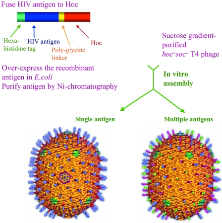
FIG. 1.
Schematic of the phage T4 in vitro assembly system. Phage T4 capsid cryo-electron microscopy reconstructions (7) are shown with antigen “spikes” artificially fused to Hoc subunits. The left reconstruction shows blue spikes representing the display of single antigen (p24-gag in the present study), and the right reconstruction shows blue, green, and pink spikes representing the display of three antigens (p24-gag, Nef, and gp41 C-trimer in the present study). Hoc subunits are shown in dark red; the hexameric gp23* protrusions and the Soc subunits bridging the gp23* subunits form the capsid shell shown in gold; in one of the hexagons of the icosahedral face (left reconstruction), the gp23* and Soc subunits are shown in blue and purple, respectively, to distinguish these subunits on the capsid lattice. The vertex at the base of the capsid represents the unique portal vertex to which the neck and tail attach (not shown).

