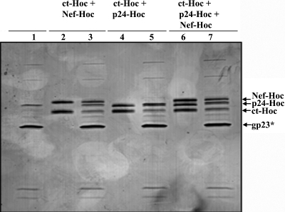
FIG. 6.
Multiple HIV antigen display. The hoc−soc− particles (1010 PFU) were incubated with two HIV antigens (ct-Hoc and Nef-Hoc [lanes 2 and 3] and ct-Hoc and p24-Hoc [lanes 4 and 5]) or three HIV antigens (ct-Hoc, p24-Hoc, and Nef-Hoc [lanes 6 and 7]) under standard in vitro assembly conditions. The samples were electrophoresed on a SDS-13% PAGE gel and stained with Coomassie blue. Lanes: 1, control hoc−soc− phage; 2, 4, and 6, combinations of the proteins as indicated at the top; 3, 5 and 7: phage showing the displayed antigens. The positions of gp23*, ct-Hoc, p24-Hoc, and nef-Hoc are labeled with arrows.

