FIG. 7.
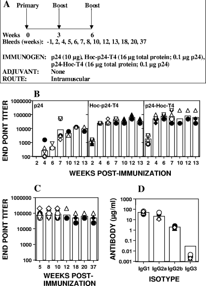
Immunogenicity of T4-displayed HIV p24. (A) Experimental design for immunization. Female BALB/c mice (six/group) were immunized by intramuscular injections at weeks 0, 3, and 6 with the antigen doses as indicated. Three mice from each group were euthanized at week 7 for analysis of the cellular responses. (B) In the first experiment, blood was collected from each of the six mice at weeks 2, 4, 6, and 7 and thereafter from the three remaining mice at weeks 10, 12, and 13. (C) In the second experiment, blood was collected from all six mice at weeks 5, 8, and 10 and thereafter from the three remaining mice at weeks 12, 18, 20, and 37. (B) Display of p24 on phage T4 enhances antibody responses to p24. BALB/c mice were immunized with p24 (10 μg), Hoc-p24-T4 (16 μg of phage protein displaying 100 ng of p24), and p24-Hoc-T4 (16 μg of phage protein displaying 100 ng of p24). Individual serum samples were analyzed in triplicate by ELISA for the presence of p24-specific IgG antibodies using baculovirus-expressed p24 as the coating antigen. Each bar represents the geometric mean endpoint titers for each group. Each symbol represents the endpoint titer for an individual mouse. (C) Display of p24 on phage T4 induces long-lasting p24-specific IgG antibodies. The experimental design is the same as in panel B except that sera were collected for up to 37 weeks and analyzed for p24-specific IgG antibodies by ELISA. (D) Subclass analysis was performed on individual serum samples collected at week 8 from p24-Hoc-T4-immunized mice (see panel C) using peroxidase-conjugated goat anti-mouse subclass specific IgG as the secondary antibody. The absorbance (A405) was measured at the 30-min endpoint. The amount of antibody in each sample was calculated from a standard graph. Each bar represents the geometric mean for each subclass, and individual serum samples from six mice are represented by the various symbols.

