FIG. 1.
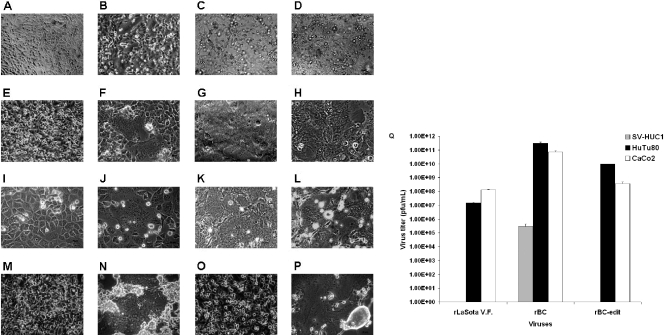
NDV is cytolytic to human tumor cells and noncytolytic in normal human cells. Shown are CPE induced by rNDV in chicken embryo fibroblast and human tumor cells. DF1 chicken embryo fibroblast cells and human tumor cell lines were either mock infected or infected with rLaSota V.F., rBC, or rBC-Edit strains of NDV at an MOI of 0.01. CPE in the form of cell fusion, syncytium formation, rounding, and destruction of the monolayer in different cells are shown. (A and B) Mock-infected and rBC-Edit-infected HEpG2 cells. (C and D) Mock-infected and rBC-Edit-infected HT1080 cells. (E and F) Mock-infected and rBC-Edit-infected PC3 prostate cancer cells. (G and H) Mock-infected and rBC-Edit-infected CaCo2 colon cancer cells. (I and J) Mock-infected and rBC-Edit-infected HuTu80 intestinal epithelial cells. (K and L) Mock-infected and rBC-Edit-infected DF1 chicken embryo fibroblast cells. (M and N) Mock-infected and rBC-Edit-infected 2fTGH human fibrosarcoma cells. (O and P) Mock-infected and rBC-Edit-infected U3A human fibrosarcoma cells. Magnification, ×40. (Q) SV-HUC1 uroepithelial cells were either mock infected or infected with rLaSota V.F., rBC, or rBC-Edit strains of NDV at MOIs of 0.01, 1.0, or 10. Culture supernatants were assayed for virus content by a plaque assay in DF1 cells at 48 h postinfection, and results of virus titer determination at an MOI of 0.01 were compared with those for viruses assayed under similar conditions in HuTu80 and CaCo2 cells. Results represent mean values + SEM from two independent experiments.

