FIG. 5.
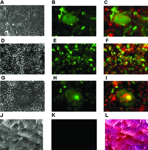
Disruption of mitochondrial membrane potential in rBC-EGFP-infected tumor cells was examined by staining with DAPI and MitoTracker Red CMX-Ros. NDV-infected cells which had a disruption of the Δψm and were undergoing apoptosis were shown by the diffuse cytoplasmic pattern of CMX-Ros with condensed chromatin. Tumor cells were infected with rBC-EGFP virus, treated 24 h postinfection with MitoTracker Red CMX-Ros for 2 h, and fixed later. Syncytium formation, EGFP expression, and mitochondrial membrane disruption in rBC-EGFP-infected cells are shown. (A) CaCo2 colon carcinoma cells, bright field; magnification, ×40. (B) Epifluorescence microscopy, ×40. (C) Diffuse staining of cytoplasm with MitoTracker Red merged with fluorescent image, ×40. (D) HEpG2 hepatocarcinoma cells, bright field, ×40. (E) Epifluorescence, ×40. (F) Diffuse cytoplasmic staining of MitoTracker Red with EGFP expression, ×40. (G) PC3 prostate cancer cells, bright field, ×40. (H) Epifluorescence, ×40. (I) Diffuse cytoplasmic staining of MitoTracker Red with EGFP expression, ×40. (J) HuTu80 cells, uninfected control cells, bright field, ×40. (K) Epifluorescence, ×40. (L) Punctuate cytoplasmic staining with MitoTracker Red, ×40.

