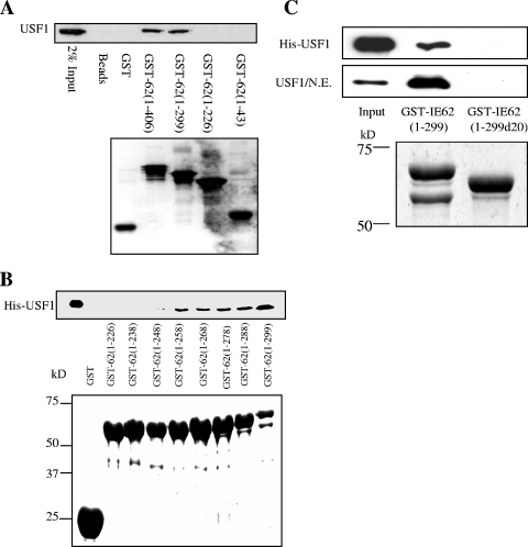
FIG. 4.
GST pull-down analysis of the region of IE62 that interacts with USF1. (A) Preliminary mapping of the region of IE62 that interacts with USF. Truncated GST-tagged IE62 proteins were coupled to glutathione Sepharose beads and incubated with nuclear extracts of MeWo cells. GST alone was used as a control. The binding of USF to the GST-IE62 fusions was examined by Western blotting (upper panel). The lower panel shows a Western blot assay using anti-GST antibody documenting the levels of the GST and GST-IE62 fusion proteins eluted from the beads. (B) Fine mapping of the USF binding region of IE62 using a series of progressive 10-amino-acid C-terminal deletions of GST-IE62 (1-299). The upper panel shows an immunoblot analysis of the level of purified HIS-USF1 binding. The lower panel is a Coomassie blue stain showing the levels of the GST-IE62 fusions eluted from the glutathione beads. (C) Binding of recombinant full-length USF1 and USF1 present in MeWo cell nuclear extracts to GST-IE62 (1-299) and GST-IE62 (1-299d20). The top two panels are an immunoblot analysis of bound USF1. The bottom panel is a Coomassie blue stain showing the levels of the two fusion proteins eluted from the glutathione beads.

