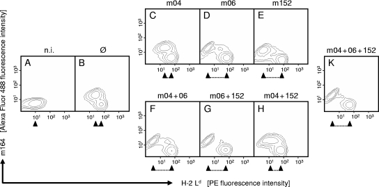
FIG. 1.
Regulation of MHC class I cell surface expression by vRAPs. Primary BALB/c-derived MEFs were either left uninfected (n.i., no infection) (A) or were infected with mutant mCMV-Δm04+06+152 lacking vRAPs (Ø) (B), with mCMV-WT expressing all three vRAPs (K), or with vRAP gene deletion mutants expressing individual vRAPs and combinations of vRAPs as indicated (C to H). Contamination of MEFs with CD11b+ cells (e.g., macrophages) was below the detection limit of cytofluorometric analysis. Two-color cytofluorometric analysis was performed after 16 h in the late early phase of viral gene expression. Contour plots represent fluorescence intensity levels for ∼35,000 live cells analyzed with no further gating. Ordinate fluorescence data represent expression of the viral cytoplasmic early-phase protein m164 (gp38/50), and abscissa fluorescence data represent cell surface expression of the MHC class I molecule Ld. Upregulated Ld expression in uninfected cells present within the infected cultures served as an internal standard. The regulating effect of vRAPs is highlighted by a caliper rule symbol. PE, phycoerythrin.

