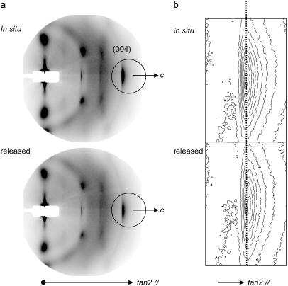
FIGURE 4.
A typical diffraction patterns (a) obtained from P. euramericana tension wood in situ and after the stress release from the first cycle of synchrotron x-ray experiment (JASRI proposal No. 2003B0463-NL2a-np). The distance between sample and camera was ∼320 mm. As in enlarged contour plot (b) of the encircled area in a, the peak position of (004) diffraction shifted toward the higher angle (to the right), indicating that the fiber repeat distance is contracted upon the release of the maturation stress. The shift was, however, in the range of a few pixels so that the longer camera length is requisite to improve the resolution of the experiment. Yet no quantitative values were obtained during this experiment but the shift was constantly observed in the same direction. Fiber axis is horizontal.

