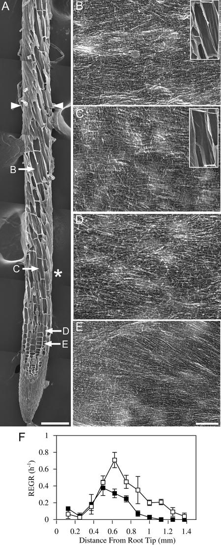
Figure 4.
After 8 h at 29°C, Cellulose Microfibrils in mor1-1 Roots Remain Predominantly Transverse to the Root Long Axis, Despite Left-Handed Twisting and Reduced Elongation Rates.
(A) Field emission scanning electron micrograph of a 5-day-old mor1-1 root tip after 8 h of growth at 29°C. The boundary of the elongation zone (arrowheads) and the points from which the higher magnification images ([B] to [E]) were taken are indicated. Left-handed cell file twisting initiates in the late elongation zone, after the point at which maximum relative elemental growth rates were recorded (asterisk). Bar = 100 μm.
(B) to (E) Higher magnification micrographs showing detailed cellulose microfibril arrangement in cells from the late elongation zone (B), near the point of maximum relative elemental growth rate (C), in the early elongation zone (D), and in the cell division zone (E). Microfibril orientation remains transverse to the root's long axis. Insets in (B) and (C) illustrate how the microfibril orientation is not perpendicular to the long axis of the twisting cell files. Bar = 300 nm.
(F) Relative elemental growth rates (REGR) measured over a 1-h period after 7 h at 29°C for mor1-1 (closed squares) and wild-type (open squares) root tips. Compared with those of the wild type, mor1-1 elongation rates are lower and the elongation zone is shorter.

