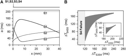
FIGURE 5.
Induction of dispersion of refractoriness by multiple extrasystoles in homogeneous 1D cable from the kinematic model (Eq. 3). (A) APD distribution in space for the S1, S2, S3, and S4 beats, which were applied at the same end of the cable. ΔTS1S2 = 211 ms, ΔTS2S3 = 65 ms, and ΔTS3S4 = 125 ms. (B) The vulnerable window w (shaded area) for the S5 beat versus the S3S4 interval for ΔTS1S2 = 211 ms and ΔTS2S3 = 65 ms. Inset shows w for ΔTS1S2 = 211 ms and ΔTS2S3 = 200 ms.

