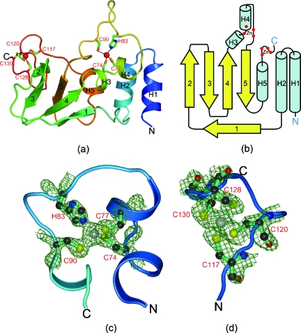
FIG. 2.
Structure of SARS-CoV nsp10. (a) Ribbon diagram of SARS-CoV nsp10 showing the arrangement of helices and strands. The secondary structures are colored from blue (N terminus) to red (C terminus) and are numbered from H1 to H5 for helices and 1 to 5 for the β-strands. (b) Topology diagram showing the connectivities between the secondary structural elements in the nsp10 structure. Helices are in cyan and strands are in yellow, with the same numbering scheme as that described for panel a. (c) Electron density observed at the first Zn2+-binding site. The residues coordinating the Zn2+ ion are shown as balls and sticks. (d) Electron density observed at the second Zn2+-binding site. The four cysteine residues coordinating the metal ion at the second Zn2+ ion near the protein C terminus are shown as balls and sticks. The 2Fo-Fc maps are contoured at 1.0 σ, where Fo and Fc are the observed and calculated structure factors, respectively.

