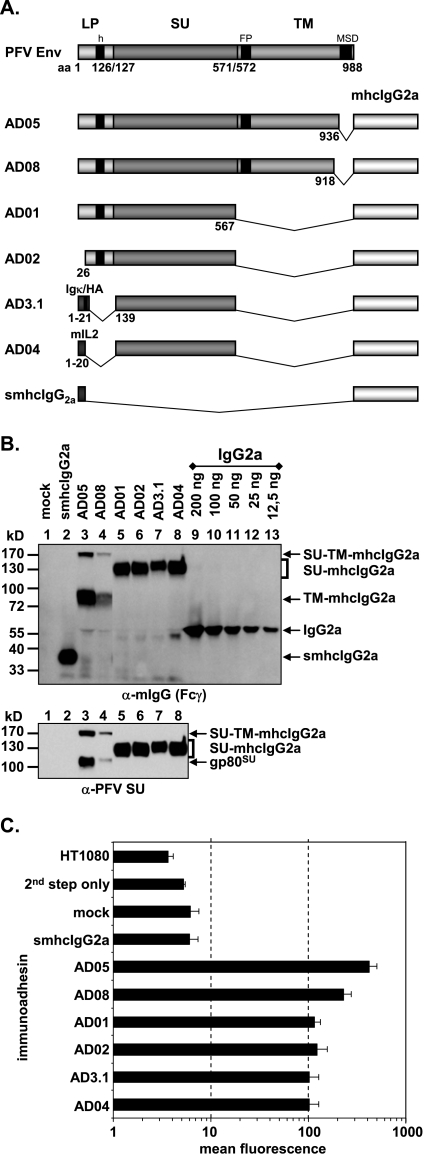
FIG. 2.
Analysis of different subdomain constructs. (A) Schematic outline of the PFV Env domain structure and the PFV Env immunoadhesins containing different domains as indicated. LP, leader peptide; SU, surface; TM, transmembrane; h, hydrophobic domain of LP; FP, fusion peptide; MSD, membrane-spanning domain; mhcIgG2a, mouse IgG2a heavy chain constant domains; IgΚ, mouse IgG kappa light chain signal peptide; mIL2, mouse interleukin 2 signal peptide; HA, hemagglutinin A epitope tag. (B) Western blot analysis of 293T supernatants (100 μl) containing different PFV Env immunoadhesins, controls, or IgG2a standard for protein concentration determination as indicated using polyclonal anti-mouse IgG-Fcγ or monoclonal anti-PFV SU-specific antibodies. The identities of the individual proteins are given on the right. (C) Mean fluorescence and corresponding standard deviation (n = 3) of different immunoadhesins and controls on HT1080 target cells. Staining was done using 50 ng immunoadhesin or control in a volume of 830 μl.

