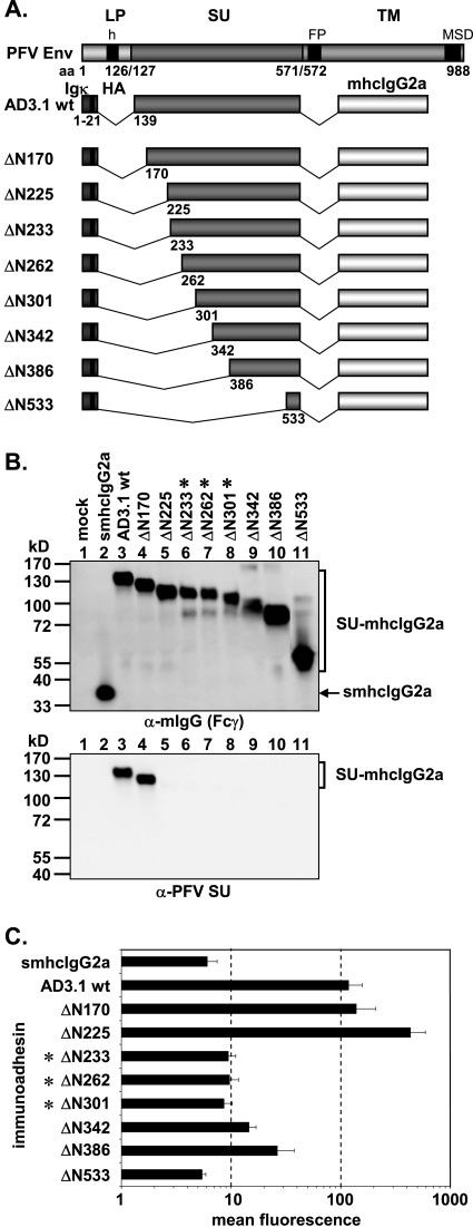
FIG. 3.
Analysis of different N-terminal truncation mutants. (A) Schematic outline of the PFV Env domain structure and the N-terminal PFV Env LP-SU immunoadhesin deletion mutants. For abbreviations, see legend to Fig. 2. (B) Western blot analysis of 293T supernatants (50 μl) containing different PFV Env immunoadhesins or purified immunoadhesins (marked by asterisks) and controls, as indicated, using polyclonal anti-mouse IgG-Fcγ or monoclonal anti-PFV SU-specific antibodies. The identities of the individual proteins are given on the right. (C) Mean fluorescences and corresponding standard deviations (n = 3) of different immunoadhesins and controls on HT1080 target cells. Staining was done using 100 ng immunoadhesin or control in a volume of 450 μl.

