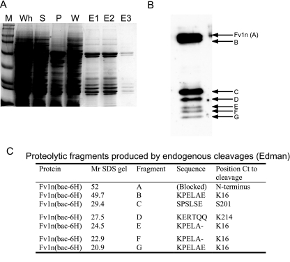
FIG. 1.
Analysis of the expression, purification, and in vivo proteolysis of Fv1n in recombinant baculovirus-infected insect cells. (A) Coomassie brilliant blue-stained SDS gel of nickel-chelate affinity purification. M, molecular weight markers; Wh, whole-cell extract; S, soluble fraction; P, pellet fraction; W, wash; E1 to E3, fractions from imidazole elution. (B) Western blot showing purified Fv1n (A) and copurifying fragments (B to G). (C) Table of the N-terminal sequences determined by automated Edman degradation. Fragments and Fv1n are labeled as in panel B. Relative molecular weights (Mr) are in thousands. Ct, C-terminal.

