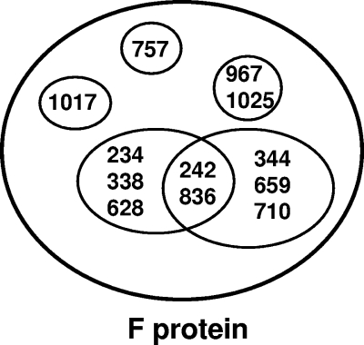
FIG. 1.
Depiction of the epitopes recognized by the hMPV F protein-specific monoclonal antibodies. Each circle represents an individual epitope on hMPV F, with the MAb binding to that epitope shown inside the circle. MAb numbers inside the intersection of circles are those monoclonal antibodies that have recognition sites comprised of a portion of two epitopes.

