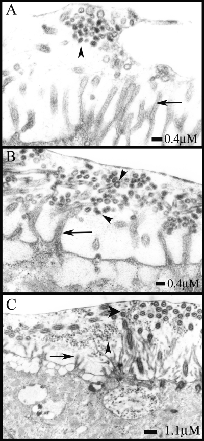
FIG. 7.
HAE cultures were infected with avian (A/Duck/Singapore/5/97) and human (A/England/26/99) influenza A viruses at an MOI of 0.1 and fixed at 12 h postinfection with perfluorocarbon-osmium tetroxide that preserves the air-surface microenvironment of HAE. Sections were processed for electron microscopy and analyzed. For the avian influenza strain, only one small cluster of budded virus particles was visible after many fields were scanned (A). Released human influenza virus particles were readily detected above the surfaces of cells. The released virus morphology was predominantly spherical (B), and virions were found to be accumulated at the level of microvillar tips (C). Scale bars are shown for each panel. Arrows, microvilli; small arrowheads, virus particles; large arrowhead in panel C, cilia.

