FIG. 1.
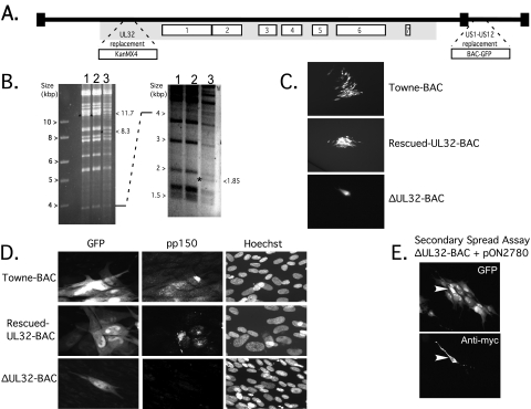
UL32-BAC structure and complementation. (A) Schematic of the HCMV TownevarATCCshort strain, Towne-BAC-derived ΔUL32-BAC genome, with the locations of the BAC-GFP insertion replacing US1 to US12 and the KanMX4 insertion replacing the entire betaherpesvirus-conserved UL32 ORF (13) indicated by expanded regions. The numbered open boxes indicate the locations of herpesvirus core genes, and the gray shaded area indicates the location of the betaherpesvirus-conserved genes (35, 36). (B) Ethidium bromide-stained images of electrophoretically separated (larger fragments, left; smaller fragments, right) HindIII restriction digests of 5 μg of Towne-BAC (lane 1), rescued-UL32-BAC (lane 2), or ΔUL32-BAC (lane 3). The 11.7-kbp fragment present in Towne-BAC or rescued-UL32-BAC is replaced by 8.3- and 1.85-kbp fragments in ΔUL32-BAC, as indicated by an asterisk on the left of the DNA fragment and by the sizes on the right side of each panel. (C) GFP expression following transfection of HFFs with rescued-UL32-BAC (a focus is shown at ×50 magnification), Towne-BAC (a focus is shown at ×50 magnification), and ΔUL32-BAC (a single cell is shown at ×100 magnification) at day 10 posttransfection. (D) GFP expression following transfection (as described for panel C) with Towne-BAC, rescued-UL32-BAC, and ΔUL32-BAC. Detection of GFP, pp150 antigen, and DNA (Hoechst) was performed on day 8 posttransfection (×300 magnification). A merge is shown to localize the pp150 accumulation. (E) Detection of GFP (top panel) and myc-tagged pp150 (bottom panel) in a secondary spread assay at 8 days following cotransfection of HFFs with ΔUL32-BAC and WT pp150 expression plasmid (pON2780) (×100 magnification). The arrowhead identifies the myc epitope-tagged pp150-positive cell.

