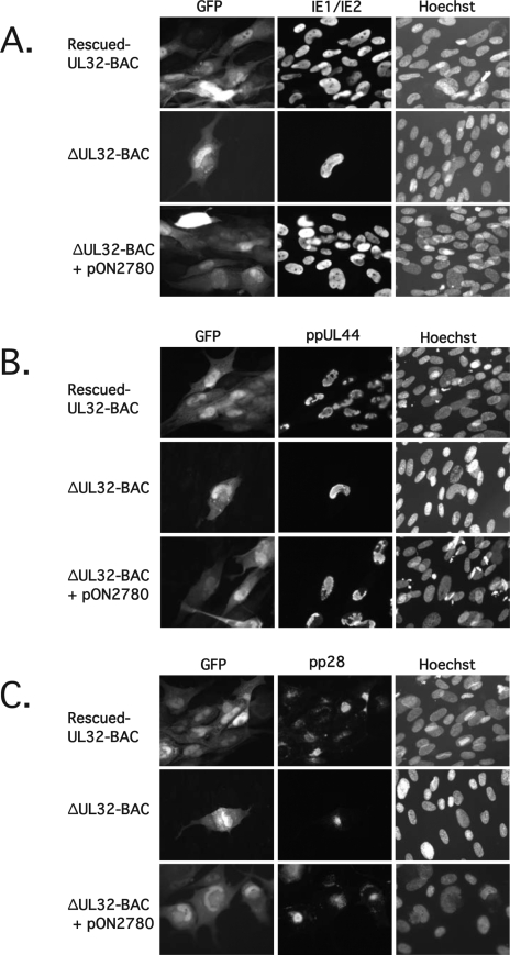
FIG. 3.
Expression of IE, early, and late viral antigens by ΔUL32-BAC. HFFs were transfected with rescued-UL32-BAC, ΔUL32-BAC, or ΔUL32-BAC plus WT pp150 expression plasmid pON2780. (A) Immunofluorescent analysis of cells fixed 8 days posttransfection and stained with FITC-conjugated MAb 810 to detect IE1/IE2. (B) Immunofluorescent analysis of cells fixed 8 days posttransfection and stained with an HCMV ppUL44 MAb followed by detection with Alexa Fluor 594-conjugated secondary antibody. (C) Immunofluorescent analysis of cells fixed 10 days posttransfection and stained with HCMV pp28 (UL99) MAb, followed by secondary detection with Alexa Fluor 594-conjugated secondary antibody. Hoechst staining was used to localize nuclear DNA. Magnification, ×300.

