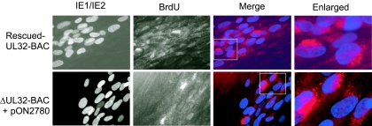
FIG. 4.
Localization of newly synthesized viral DNA. (A) Immunofluorescent images of HFFs transfected with rescued-UL32-BAC (top panels) or ΔUL32-BAC plus WT pp150 (bottom panels), followed by a BrdU (10 μM) pulse for 12 h and a 48-h chase. BrdU was localized with an Alexa Fluor 594-conjugated MAb. A false color merge was used to localize BrdU-stained viral DNA (red) in a juxtanuclear position relative to IE1/IE2-positive nuclei (blue). The merged images contained in boxes are enlarged in column 4. Cells were fixed in methanol:acetic acid (3:1). Magnification, ×400 for columns 1 to 3 and ×1,000 for column 4.

