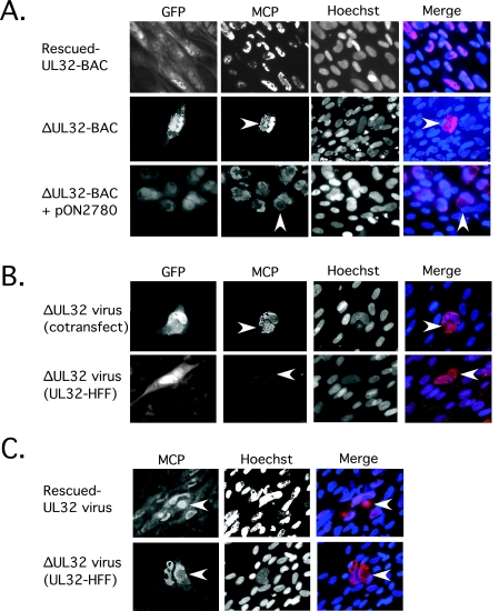
FIG. 5.
Localization of MCP within the cytoplasm of ΔUL32-BAC-transfected or -infected cells. (A) Immunofluorescent images of HFFs fixed with 3.7% formaldehyde at day 10 following transfection with rescued-UL32-BAC (top panels), ΔUL32-BAC (middle panels), and ΔUL32-BAC plus WT pp150 (bottom panels). (B) Immunofluorescent images of HFFs infected with supernatant virus from ΔUL32-BAC plus WT pp150-cotransfected cells [ΔUL32 virus (cotransfect)] fixed on day 10 postinfection (top panels) and HFFs infected with supernatant virus recovered from the ΔUL32-BAC-transfected pp150-expressing HFF line [ΔUL32 virus (UL32-HFF)] fixed at day 7 postinfection (bottom panels). (C) Immunofluorescent images of MCP accumulation at day 8 postinfection with rescued-UL32 (top panels) and ΔUL32 (bottom panels) virus fixed with methanol:acetic acid (3:1). GFP-positive cells are shown in the far left panel for all samples fixed with 3.7% formaldehyde (methanol:acetic acid fixation destroys GFP detection). All staining for MCP was done with MAb 28-4 along with Alexa Fluor 594 secondary antibody. Hoechst was used to stain nuclei, and a false color merge with MCP antigen localization is shown. The arrowhead indicates cytoplasmic localization of MCP. All images were collected with equivalent exposure times. Magnification, ×200.

