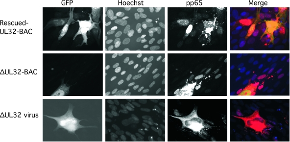
FIG. 7.
Localization and distribution of pp65 in ΔUL32-BAC-transfected and ΔUL32 virus-infected cells. Immunofluorescent images of rescued-UL32 (top panels) or ΔUL32 (middle panels) transfection at day 10 posttransfection compared to ΔUL32 virus infection (bottom panels) at day 7 postinfection. Detection of pp65 was accomplished with MAb 28-19 followed by Alexa Fluor 594 secondary antibody. Magnification, ×400.

