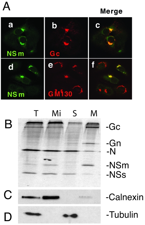
FIG. 2.
Intracellular localization and determination of the membrane integrality of NSm. (A) Colocalization of NSm with Gc (panels a to c) and Golgi matrix protein GM130 (panels d to f). wt BUNV-infected BSR-T7/5 cells were stained with a mixture of anti-NSm antibody and either anti-Gc MAb 742 or anti-GM130 MAb. NSm stains green (panels a and d), and Gc and GM130 stain red. Merged confocal microscopic images are also shown, with colocalization shown in yellow (panels c and f). (B) NSm is an integral membrane protein. Vero E6 cells were infected with wt BUNV and radiolabeled with [35S]methionine, and membrane fractions were prepared as described in Materials and Methods. Total (T) and microsomal (Mi) fractions were collected, and membranes were extracted with sodium carbonate to yield supernatant (S) and membrane (M) samples. The fractions were analyzed by SDS-PAGE. The positions of viral proteins are indicated at the right. (C). Western blot analysis of the gel using anti-calnexin antibodies as a marker for membranes. (D) Western blot analysis of the gel using anti-tubulin antibodies as a marker for the cytosolic fraction.

