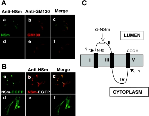
FIG. 3.
Determination of the topology of BUNV NSm. Vero E6 cells were infected with wt BUNV (A) or recombinant virus rBUNM-NSm-EGFP (B) and semipermeabilized by the freeze-thaw technique. Cells shown in the upper row of each set were further permeabilized with 0.2% Triton X-100-PBS (panels a to c in each case). Before examination by confocal microscopy, the wt BUNV-infected cells were costained with rabbit anti-NSm serum and anti-GM130 MAb and the rBUNM-NSm-EGFP-infected cells were stained only with anti-NSm serum. In panel A, NSm stains green and GM130 stains red, and in panel B, the EGFP autofluorescence of NSm-EGFP protein shows as green and the NSm antibody stains red. Merged confocal images are also shown. NSm antibodies can only react with NSm or NSm-EGFP in fully permeabilized cells. (C) The predicted topology of NSm. Hydrophobic domains are shown as black columns across the intracellular membrane. The positions of the predicted domains I to V and of the anti-NSm epitope are marked.

