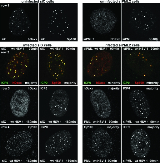
FIG. 6.
ICP0 initially colocalizes with and then disperses Sp100 and hDaxx in both siC and siPML2 cells. siC and siPML2 cells were infected with wt HSV-1 (MOI of 2), and samples were analyzed by immunofluorescence after 90 and 180 minutes of infection. The panels are appropriately labeled to indicate the cell type, the time point, and the staining regimen used. Row 2 shows individual cells dual labeled for the indicated proteins. Rows 1, 3, and 4 show the separated red and green channels for either single siC cells (left of panels) or siPML2 cells (right of panels). Row 1 shows control uninfected siC and siPML2 cells, and rows 2 to 4 show infected siC and siPML2 cells as indicated.

