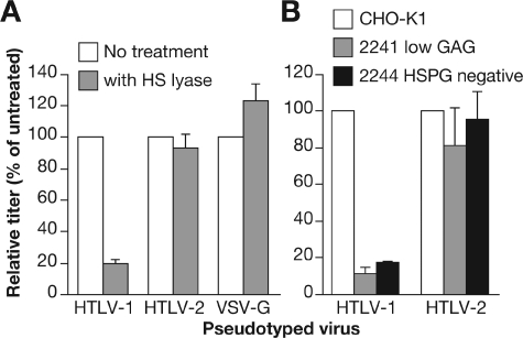
FIG. 2.
Effects of HSPG cell surface expression on HTLV-1 Env and HTLV-2 Env-mediated entry. (A) COS-7 cells (106) were suspended in 200 μl of HS lyase buffer (20 mM Tris, pH 7.4, 0.01% BSA, and 4 mM CaCl2) and then incubated for 2 h at 37°C with either 120 mU of HS lyase (gray bars) or left untreated (white bars). HTLV-1 Env-, HTLV-2 Env-, or VSV-G-pseudotyped virus particles were generated and used to transduce the COS-7 cells without spinoculation, and the titers were determined 3 days later. (B) HTLV-1 Env- and HTLV-2 Env-pseudotyped virus particles were used to transduce CHO-K1 cells (white bars), CHO-K1 2241 cells (gray bars), and CHO-K1 2244 cells (black bars) without spinoculation, and the titers were determined 3 days later. The data are the average of two independent experiments; error bars represent standard deviation. The titer for each of the pseudotyped virions on COS-7 cells in the absence of treatment (A) was as follows in experiment 1: HTLV-1, 6.8 × 10³; HTLV-2, 5.4 × 10⁴; VSV-G, 4.2 × 10⁴. In experiment 2 the titers in COS-7 cells were as follows: HTLV-1, 5.9 × 10⁴; HTLV-2, 3.7 × 10⁴; VSV-G, 7.2 × 10⁴. The titer for each of the pseudotyped vectors on the parental CHO-K1 cells (B) in experiment 1 were 4.4 × 10³ for HTLV-1 and 4.0 × 10³ for HTLV-2. In experiment 2 the titers in CHO-K1 cells were 7.8 × 10⁴ for HTLV-1 and 5.7 × 10⁴ for HTLV-2. Data shown are representative; an additional two (A) or three (B) experiments were performed.

