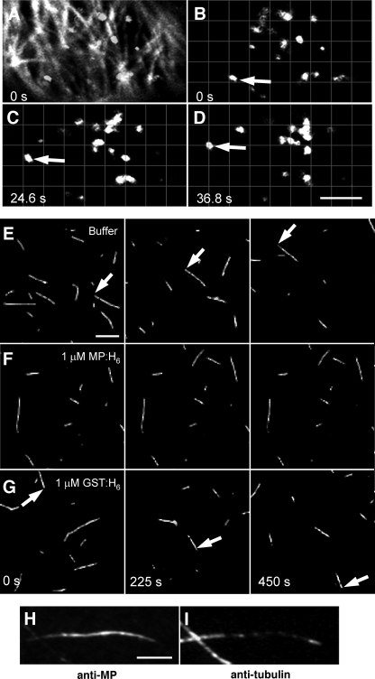
FIG. 7.
Effects of MP MAP activity on molecular motor transport. (A to D) The microtubule association of MP does not inhibit Golgi stack movements in BY-2 cells infected with TMV-MPΔC55-GFP or ectopically expressing MPΔC55-GFP. In cells where MPΔC55-GFP is clearly associated with microtubules (panel A, overlay of the MPΔC55-GFP signal [filaments] and Man-1-mRFP signal [bodies, i.e., Golgi complexes], compare with panel B), movements of Man-1-mRFP-labeled Golgi stacks (B to D) are apparent at an average velocity of 0.095 ± 0.031 μm/s. Bar, 4 μm. (E to G) Time-lapse recordings of fluorescent microtubules translocating along the surface of a perfusion chamber coated with kinesin-1. Bar, 4 μm. (E) In the absence of exogenous proteins, microtubules translocate at an average velocity of 0.2 ± 0.052 μm/s. (F) The presence of 1 μM MP-H6 completely abolishes kinesin-dependent microtubule translocation. (G) Kinesin-dependent microtubule movements are still apparent in the presence of 1 mM GST-H6 at an average velocity of 0.14 ± 0.058 μm/s. (H and I) Microtubules immobilized within kinesin-coated perfusion chambers are highly decorated in the presence of 1 μM MP-H6. Bar, 1.5 μm.

