
FIG. 5.
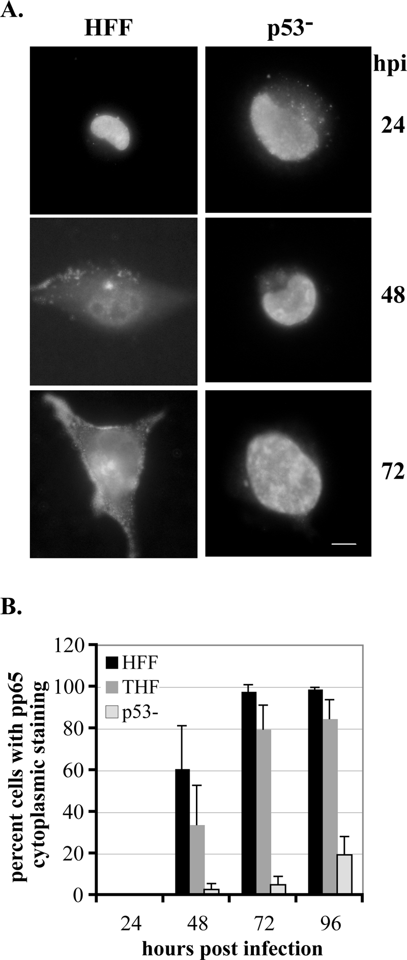
pp65 relocalization is impaired in p53−/− cells. Cells were seeded onto glass coverslips and infected, and coverslips were harvested at the indicated times p.i. Cells were stained for the presence of pp65 as described in Materials and Methods. Cells were scored for localization of pp65 within the cell as delineated in the text. (A) Representative staining for pp65 in HFF and p53−/− cells at the indicated times p.i. Images depict pp65 localization, with HFF images showing the three phases described in the text. Localization begins solely in the nucleus, gradually moves into the cytoplasm at 48 h p.i., and is almost completely in the cytoplasm by 72 h p.i. in these wt cells. For the purposes of our experiments, cells were scored as positive for pp65 in the cytoplasm if they progressed to the stage portrayed at 48 h p.i. in HFF cells. (B) Graphic representation of the results of scoring at least three experiments for each cell type for movement of pp65 into the cytoplasm. At least 100 cells were scored for each cell type at each time point in each experiment. Error bars represent 1 standard deviation.