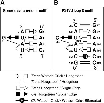
FIG. 3.
Tertiary-structural model of PSTVd loop E. (A) Paradigmatic sarcin/ricin motif based on X-ray crystal structures (adapted from reference 29 with permission from Elsevier). (B) Inferred PSTVd loop E structural model. The dashed arrows indicate local changes in the strand orientation. All symbols that denote non-Watson-Crick base pairs and strand orientations are based on a report by Leontis and Westhof (34). Circles, squares, and triangles indicate the participation of Watson-Crick, Hoogsteen, and sugar edges, respectively. Open symbols indicate base pairs with a trans orientation of the glycosidic bonds, and closed symbols indicate base pairs with a cis orientation.

