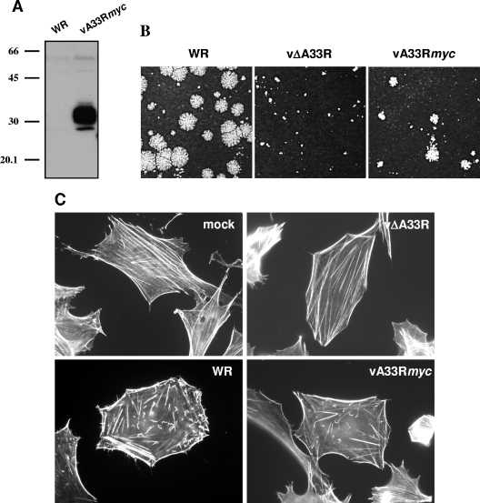
FIG. 2.
Characterization of a recombinant vaccinia virus expressing a myc-tagged version of the A33 protein. (A) Analysis of A33myc expression by Western blotting. Extracts of BSC-1 cells infected with the recombinant vaccinia virus expressing fusion protein A33myc (vA33Rmyc) or control virus (WR) were probed with anti-myc-HRP antibody. Positions of protein molecular mass markers (in kDa) are indicated. (B) Plaque formation by vA33Rmyc. BSC-1 cell monolayers infected with WR, parental vΔA33R, or vA33Rmyc were incubated for 2 days, stained with crystal violet, and photographed. (C) Induction of actin tails by vA33Rmyc. BHK-21 cells infected for 7 h were fixed, permeabilized, and incubated with 1 unit/ml Alexa594-phalloidin. Note the presence of actin tails in vA33Rmyc-infected cells.

