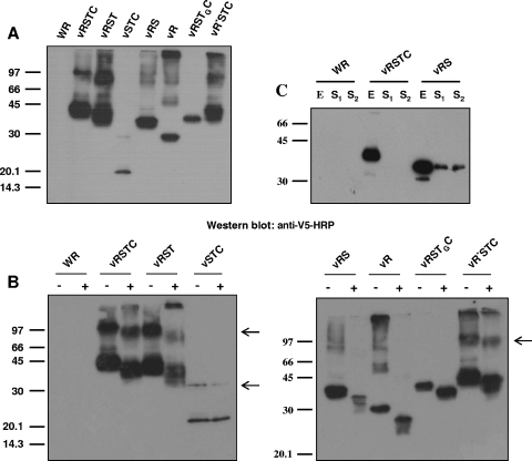
FIG. 7.
Characterization of V5-tagged, mutated B5 versions. (A) Western blot analysis. BSC-1 cells were infected with the viruses indicated at the top of the panel at five PFU per cell for 24 h. Western blots were probed with anti-V5-HRP antibody. (B) Analysis of N glycosylation. BSC-1 cells infected with the viruses indicated in the presence (+) or absence (−) of 1 μg/ml tunicamycin were harvested at 24 h postinfection. Western blots were probed with antibody against the V5 epitope conjugated with horseradish peroxidase. Arrowheads point to B5 complexes, which are absent in extracts of cells infected with viruses expressing B5 versions RS, R, and RSTGC. Positions of protein molecular-mass markers are indicated (in kDa). (C) Secretion of B5 soluble version from vRS-infected cells. BSC-1 cells were infected with the viruses indicated at five PFU per cell, harvested at 24 h postinfection, and probed with anti-V5-HRP antibody. E, total cell lysate; S1, culture medium filtered through 0.1-μm filters (to eliminate vaccinia virus) and concentrated; S2, culture medium concentrated. Note the presence of the B5 version in the supernatant of vRS-infected cells.

