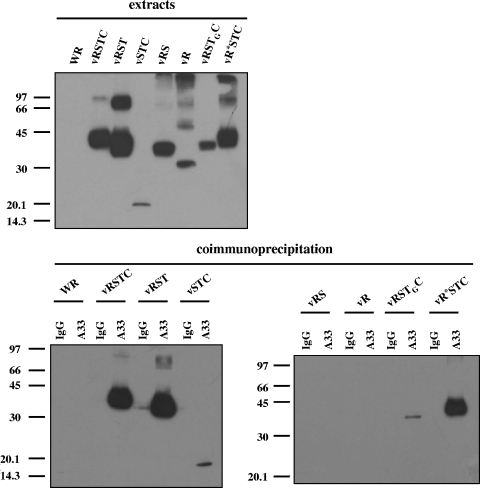
FIG. 9.
Mapping of the A33-B5 interaction site by coimmunoprecipitation. BSC-1 cells were infected with the viruses indicated above the panels at five PFU per cell and harvested at 24 h postinfection. The top panel shows infected cell extracts prior to immunoprecipitation, probed with anti-V5 antibody conjugated with horseradish peroxidase. Immunoprecipitations were performed with either control immunoglobulin G (lanes IgG) or anti-A33 antibody (lanes A33), as indicated above the lower panels. Immunoprecipitated material was resolved by SDS-PAGE and subjected to Western blot analysis with anti-V5 antibody conjugated with horseradish peroxidase. Molecular-mass markers are indicated (in kDa).

