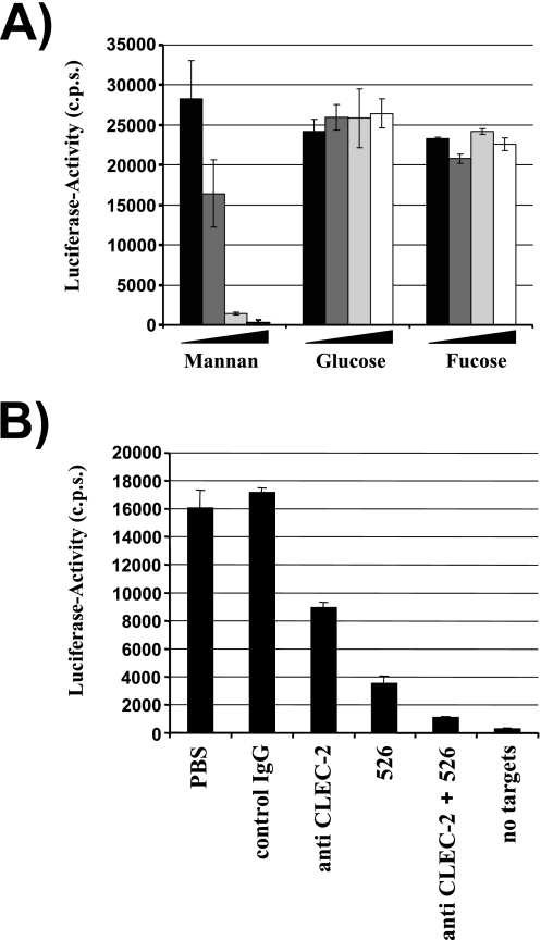
FIG. 6.
A substantial fraction of the HIV-1 capture activity of platelets is dependent on DC-SIGN. (A) The capture assay was carried out as described in the legend for Fig. 5. However, platelets were incubated with rising concentrations of mannan, glucose, or fucose before virus was added. The following carbohydrate concentrations were employed: 0 μg/ml (black bars), 5 μg/ml (dark-gray bars), 10 μg/ml (light-gray bars), and 20 μg/ml (white bars). Similar results were obtained in an independent experiment. (B) HIV-1 capture by platelets was assessed as described for panel A. However, cells were incubated with the indicated inhibitors (MAb 526 blocks ligand binding to DC-SIGN) before addition of virus. Similar results were obtained in three independent experiments. Error bars indicate standard deviations. PBS, phosphate-buffered saline; IgG, immunoglobulin G; c.p.s., counts per second.

