FIG. 3.
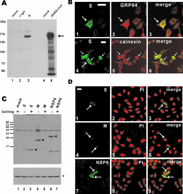
Expression of SARS-CoV proteins in cultured cells. (A) Western blot analysis of S protein expression. pLenti-β-gal-transfected and pLenti-S-transfected 293FT cells (lanes 2 and 3) and SARS-CoV-infected FRhk4 cells (lane 5) were lysed and immunoblotted with rabbit polyclonal anti-S antibody. The arrow highlights S protein. (B) Subcellular localization of SARS-CoV S protein. HeLa cells were transfected with pLenti-S and then costained with rabbit anti-V5 and either rat anti-GRP94 (panel 2) or mouse anti-calnexin (panel 5). The S and GRP94-calnexin fluorescent signals are shown in merged images, and colocalization is shown in yellow (panels 3 and 6). Transfected cells are highlighted with arrows. Bar, 30 μm. (C and D) Expression of other SARS-CoV proteins. 293FT cells were transfected with pLenti-E/M/NSP6, and protein expression was analyzed by immunoblotting (C) or confocal immunostaining (D) with rabbit polyclonal anti-V5 antibody. To prevent thermal aggregation of M and NSP6 (28), protein samples were not heated before being loaded onto the SDS-polyacrylamide gel electrophoresis gel. Nuclear morphology of cells was visualized by propidium iodide (PI) staining. In panel C, the arrowheads highlight E/M/NSP6 proteins and the asterisk indicates the control protein α-tubulin.

